


1917/1918 Spanish Flu & Encephalitis lethargica - resembles 'Covid19' and 'Long Covid'
'Sleepy encephalitis' - my nickname - positive feedback loop, hyperinflammation is another nickname.

John Cullen is suggesting that what was really going around in greater severity during the “CoV Pandemic” was a GoF flu virus dating back to 1917. Timeline, it would have been part of the 1918 ‘Spanish Flu’ epidemic, but seems to have been a slightly different version with a particular adverse effect appearing later in some people called Encephalitis lethargica (EL). EL is a type of encephalitis, brain inflammation, which caused symptoms of sleepiness, lethargy. The changes observed in brain tissue are similar sounding to ‘LongCovid’ or ‘Covid19’. John Cullen has a Thread with several videos about the pandemic and odd disappearance of flu - first post of his Thread. Was Fauci covering up flu GoF research by trying to make it seem as if everyone was getting ‘CoV’ instead of a bad flu?
Health Solutions summary - whatever we want to call it - hyperinflammation or cytokine storm or brain inflammation - STOPPING the positive feedback loop of hyperinflammation is a major need. (Pom paper abstract post)
Thiamine! niacin! antioxidants! Vitamin D3 and K2, magnesium! zinc! resistant starches! Nrf2 promoters/NF-kB inhibitors like niacin and pomegranate peel and other functional foods and phytonutrients and omega 3 fatty acid. Tryptophan and arginine, amino acids, might be helpful. Lysine adequacy and glycine and cysteine would also help - protein adequacy in general, including red meat for taurine and vitamin A metabolism would likely be helpful. BUT avoid glyphosate and support the gut microbiome and mitochondria.
More difficult - don’t be young, don’t be male, and/or try to avoid doing strenuous exercise while being young and male - or add extra antioxidants and polyphenols to help support detox and immune function recovery after exercise. Marik Protocol (thiamine and vitamin C) can help cytokine storm/sepsis, if it isn’t banned by the FDA or local hospital….
Research review
The unusual illness may be a signature of sorts. Is ‘Covid19’ and ‘Long Covid’ maybe really a bad flu followed by a ‘sleepy encephalitis’ later? Brain fog and extreme tiredness would be symptoms.
“…typical depictions of EL sleep:
During the now increasing somnolence it is not unusual to observe patients, as soon as they are left to themselves, fall asleep while sitting or standing, and even while walking or during meals, with indications of tiredness and yawning, with food in their mouths. They have the appearance of being in a light slumber or sometimes even of deep sleep with its typical breathing pattern, even snoring. If aroused by calling or shaking, they wake up quickly and completely, are well oriented and fully conscious, and can respond appropriately to questioning. They are fully aware of the situation, execute all requests promptly, get up when requested, walk about, but, if left to themselves, soon relapse into sleep. This sleep therefore appears deceptively similar to normal sleep (Economo 1929a, p 41).
Some lethargic patients state that during the whole time that they were supposed to be asleep that they could hear everything that was going on around, but could not rouse themselves. In many apparently asleep, one is surprised on asking a question to find how readily it is answered, without any apparent waking up. In other cases the reply might be delayed an appreciable time, but shows clearly that the question was understood (Hall 1924, p 73).
Sachs (1919) commented in a similar vein: “the patient lay inert with closed eyes and expressionless face but was apparently aware of what was going on about him and readily responded by nodding to questions that were put to him in a low tone of voice”. This lethargy typically lasted from a few days to a few weeks; in exceptional cases it might last for months, but in such cases it tended to progress to a deeper form, sometimes to a comatose state from which recovery was unlikely.” (Foley, 2009, II)
What follows is excerpts from two papers about EL, the unusual adverse reaction that seemed associated with the Spanish Flu, but not exactly. I then looked for similarities with EL symptoms or pathology and those observed in patients with ‘Covid19’.
T-cells and strenuous exercise
The sleeping/encephalitis associated with 1917 flu strain occurred more often in younger age groups and in males - risk groups seen with 'CoV jab' adverse reactions. T-cell immunity changes around age 10 - this was a known risk difference seen with 'CoV'. Strenuous exercise can set up a cytokine storm.
“The preferred age of onset by EL did not vary significantly throughout the 1920s, with those aged 12 to 35 years most at risk. While this age range also figured prominently in the mortality statistics for the 1918/19 influenza, lending the mortality curve its atypical W-shape, it is notable that the curves for succeeding influenza epidemics (some of which involved local mortality rates higher than those of the “great influenza”) had by 1920 resumed the usual U-shape (Collins 1945; Crome 1954). EL generally spared infants to a greater extent than did influenza (Almasio, 1921).” (Foley, 2009, II)
Male gender - more at risk for the encephalitis severe illness following flu, but not more at risk for flu compared to females.
“Finally, influenza played no favorites with respect to gender, but it was widely reported that there were up to twice as many male as female EL cases, although the prognosis in the latter tended to be worse (Stern 1936). On the other hand, the analysis of 122 United States EL cases indicated that 54% occurred without preceding influenza – but that 71% of EL cases with preceding influenza were male, while the numbers of cases without influenza were evenly divided between the genders. Curiously, the influenza attack rate in EL patients (46%) was higher than for the general American population (c. 30%; Smith 1921).” (Foley, 2009, II)
Parkinson’s Disease like developments in EL (sleepy encephalitis) and ‘Covid19’
Parkinson's Disease like symptoms in some cases following flu were linked to the EL condition by another researcher in the field. That suggests mitochondrial damage is involved, and vitamin K2 in high dose might help restore more normal function. (Vos, et al., 2012).
“Significantly, Leichtenstern’s much cited reference to “paralysis agitans” (parkinsonism) as a post-influenza complication was not part of his description of comatose influenza, but was rather included as an aside under “influenza epilepsy and other irritative motor signs”, which also included chorea, hiccough and other phenomena as after-effects of influenza (Leichtenstern 1912, p 169). Stern regarded these latter cases as “undoubtedly” being EL cases, while at the same time not committing himself to a direct causal relationship between influenza and EL (Stern 1928, p 353).” (Foley, 2009, II)
Excerpt that was in my post yesterday:
‘Vitamin K2 is a mitochondrial electron carrier that rescues pink1 deficiency.’
“Human UBIAD1 localizes to mitochondria and converts vitamin K1 to vitamin K2. Vitamin K2 is best known as a cofactor in blood coagulation, but in bacteria it is a membrane-bound electron carrier. Whether vitamin K2 exerts a similar carrier function in eukaryotic cells is unknown. We identified Drosophila UBIAD1/Heix as a modifier of pink1, a gene mutated in Parkinson’s disease that affects mitochondrial function. We found that vitamin K2 was necessary and sufficient to transfer electrons in Drosophila mitochondria. Heix mutants showed severe mitochondrial defects that were rescued by vitamin K2, and, similar to ubiquinone, vitamin K2 transferred electrons in Drosophila mitochondria, resulting in more efficient adenosine triphosphate (ATP) production. Thus, mitochondrial dysfunction was rescued by vitamin K2 that serves as a mitochondrial electron carrier, helping to maintain normal ATP production.” (Vos, et al., 2012)
If something is wiping out beneficial gut microbiome species and causing a sudden lack of vitamin K2, then that might be a factor in symptoms and explain why drinking Bubble Tea could have been protective - by supporting the beneficial species. Zinc is needed in generous amounts by those species too. The 'CoV' illness is associated with a sudden loss of Bifidobacterium, as shown by Sabine Hazan, M.D..
Bifidobacterium species are used for the commercial production of menaquinone 7 - vitamin K2, however Bacillus natto is more typically used for commercial vitamin K2 production or fermented food production = Natto, fermented soybeans. (Brave AI summary)
Foley wrote a second paper about EL and flu:
"The adage that the pathologist has the final word regarding diagnosis is particularly pertinent when considering neurological disease. The neuropathology of EL cannot be described in detail here, nor can the 1920s debate regarding what constituted “encephalitis” be pursued (review: Spatz 1930): discussion must be limited to features which distinguished EL from influenza encephalitis. Economo cited four features of EL which distinguished its neuropathology from that of influenza encephalitis:
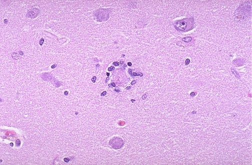
neuronophagia [Image: "neuronophagia in which a dying neuron is surrounded by microglial cells. Neuronophagia can occur in a variety of degenerative diseases, such as amyotrophic lateral sclerosis, and inflammatory conditions, such as encephalitis" (webpath.med.utah.edu)];
its preference for the grey matter of the brain;
its non-hemorrhagic character;
and the predominantly lymphocytic nature of the inflammation process.
Economo’s early reports were largely corroborated by subsequent investigators, their findings encapsulated by the Swiss neuropathologist Tobler’s comprehensive title of his 1920 paper: “acute, focal, disseminated, non-purulent, principally lymphocytic, infectious-toxic, epidemic polioencephalomyelitis”.” (Foley, 2009, III)
"The following features were consistently reported in examinations of the EL brain:
non-hemorrhagic acute inflammation of the grey matter with largely negative macroscopic findings;
superficial congestion, occasional meningeal hemorrhages;
especial involvement of substantia nigra, locus ceruleus and other brainstem areas; no Lewy bodies, but widespread neurofibrillary tangles (no plaques);
lymphocytic infiltration in basal ganglia, midbrain, pons, Sylvian aqueduct;
minor to major neuronophagia;
minor involvement of cerebral cortex and cerebellum.
(representative reports: Marinesco 1918; Bassoe and Hassin 1919; Trétiakoff 1919; Stern 1919/20; Buzzard and Greenfield 1919; Marie and Trétiakoff 1920; Siegmund 1920; Boyd 1920/21; Kuczynski and Wolff 1921; Klarfeld 1922; Agostini 1925; Greenfield 1927; Lucksch 1928; Rostan 1928; Pette 1932; Hassler 1938; Klaue 1940; Greenfield and Bosanquet 1953; reviewed: Economo 1929a; Rietti 1935; Stern 1936; see also Bernheimer et al 1973)..” (Foley, 2009, III)
The grey matter of certain regions was more likely to be damaged in EL:
“The focal damage exhibited preference for the grey substance of certain brain regions (the peri-aqueductal grey, tegmentum, hypothalamus, distal striatum and pallidum). Particularly liable to catastrophic damage was the substantia nigra, the hallmark of EL being its almost total cellular and pigment loss and replacement by a pale glial scar.” (Foley, 2009, III)
What did we see in 785 people after 'SARS-CoV-2'?
“We identified significant longitudinal effects when comparing the two groups, including (1) a greater reduction in grey matter thickness and tissue contrast in the orbitofrontal cortex and parahippocampal gyrus; (2) greater changes in markers of tissue damage in regions that are functionally connected to the primary olfactory cortex; and (3) a greater reduction in global brain size in the SARS-CoV-2 cases.” (Douaud, et al., 2022)
What might help 'Sars-CoV-2' or Parkinson's risk?
Inhibition of NF-kB. (Chaudhry, et al, 2020)
What can inhibit NF-kB?
…pomegranate peel, (Zhang, et al., 2024), niacin, citrus peel, garlic, omega 3 fatty acids, butyrate, and many other functional foods and phytonutrients that promote Nrf2, also act as inhibitors of NF-kB because a circadian clock protein is shared by the two pathways. During health, that would be like a night shift and a day shift. Modern life tends to push us into the inflammatory ‘day shift’ all of the time - all NF-kB and no clean up and repair at night be the Nrf2/Sirt3 pathway. (Nrf2 post) (Sirt3 post)


“Results: The findings revealed that PPE significantly improved the symptoms of [Diarrhea-predominant irritable bowel syndrome] IBS-D, ameliorated intestinal inflammation, and promoted intestinal barrier function. The target genes in the MAPK and NF-κB signaling pathways were significantly enriched and down-regulated. Molecular docking and Western blot assays were performed to verify that PPE had a high affinity for the protein candidates in these pathways, and significantly down-regulated the expression of p-IκB, p-p65, p-JNK, p-p38, and p-ERK1/2.” (Zhang, et al., 2024)
Pomegranate peel tends to be healing throughout the body, is very effective against cancer, and can clump nanoparticles, and is a natural inhibitor of the GLP-120 fusion cleavage protein found in HIV-1 and the chimeric spike/alleged SARS-CoV-2 virus. ….Maybe if I say that often enough, it will help more people. (Just eat some inner pith or make tea, this is not hard…)
From the first EL paper by Foley - PCR testing isn't really helpful for cerebrospinal fluid:
“Even today, influenza virus and genetic material can only rarely be isolated from cerebrospinal fluid (CSF) or autopsied brain material incases of presumed influenza CNS infection (Johnson 1998, pp 214f.; Toovey 2008). An Austrian study of influenza encephalopathy found that even highly sensitive PCR techniques could ascertain its presence in CSF in only 1 of 18 cases (Steininger et al 2003).” (Foley, 2009, II)
The PCR testing in current use wouldn’t detect the 1917 flu strain at all.
Glymphatic flow - brain clean-up - happens at night during a restful sleep. Dehydration would increase risk/decrease clean up.
From the second paper by Foley on EL - the distinguishing characteristics of the ‘sleepy encephalitis’ (my nickname for EL) is changes in grey matter near the floor of the third and fourth ventricles and aqueduct of the brain which are main points of flow into or out of the brain.
““… The current encephalitis epidemic is distinguished not through fundamental anatomical differences when compared with cerebral inflammations of other etiology, but rather the characteristic and consistent localization of the changes in the central grey, on the floor of the third and fourth ventricles, and around the aqueduct (Siegmund 1920).”” (Foley, 2009, III)
What do we see in 'Sars-CoV-2'?
"SARS-CoV-2 is known to penetrate the olfactory mucosa, causing loss of smell, and may enter the brain, migrating from the cribri-form plate along the olfactory tract2 or through vagal or trigeminal pathways; however, definitive evidence for this is lacking. SARS-CoV-2 could pass the blood-brain barrier (BBB) because inflammatory cytokines induce BBB instability or via monocytes.4 It could reach brain tissue via circumventricular organs (CVOs), midline structures around the third and fourth ventricles, that monitor blood and cerebral spinal fluid content via fenestrated capillaries lacking the junctional proteins expressed in the BBB."
- from 'How COVID19 Affects the Brain', (Boldrini, et al., 2021)
We also see Neuronophagia in ‘Covid19’, but not associated with SARS virus mRNA.... that is interesting:
"Histopathologic analysis of whole human brain showed microglial nodules and phagocytosis of neurons (neuronophagia) in brain stem and less frequently in cortex and limbic structures, associated with sparse lymphocytic infiltration, and no correlations between histopathologic findings and levels of viral messenger RNA in the same brain.5" (Boldrini, et al., 2021)
Cytokine storm, elevated TNF-alpha, and excitotoxicity are also seen in 'Covid19', 'Sars-CoV-2' infection. Interventions/treatment might include "kynurenine pathway modulators (minocycline).3" from 'How COVID19 Affects the Brain', (Boldrini, et al., 2021)
.... higher dose niacin would support or bypass dysfunction in the kynurenine pathway. (Kynurenine Pathway, Wikipedia) Pomegranate peel also supports that pathway.
Reduced serotonin and dopamine would be symptoms/side effects of a disrupted kynurenine pathway - it is NAD+ production.
A couple 2020 Tweets of mine re kynurenine being elevated in ‘Covid19’.
"concentration...kynurenine [elevated] & arginine [decreased]...could distinguish COVID-19 patients from healthy participants & other critically ill patients with 98 per cent accuracy." (schulich.uwo.ca) // Arginine is an amino acid that is depleted in sepsis: (Badurdeen, et al., 2015) — (x.com/deNutrients)
“We hope our findings can lead to faster diagnosis, identify patients most at risk of poor outcomes and targets for novel treatments,” says Dr. Douglas Fraser. His new study shows #COVID19 affects levels of specific metabolites in the blood. @WesternU, @lawsonresearch, @UAlberta; — (x.com/SchulichMedDent)
‘Research News: Biomarkers could be used in a quick, inexpensive COVID-19 blood screening tool’ (schulich.uwo.ca)
Glyphosate may be interfering with the kynurenine pathway, image via Anthony Samsel, (Samsel and Seneff, 2013) (x.com/deNutrients) - so avoid glyphosate. That isn’t easy. Buy organically grown foods, is the basic need. High risk - commercial oats and wheat, chickpeas/humus, peas, Round-Up Ready soy, other stuff. Cheerios, granola bars, many common foods… Glyphosate is also harmful to our gut microbiome and we need the good guys to make vitamin K2 and butyrate for us. Feed them zinc, resistant starches and whole foods, and try to limit processed foods which support negative species but don’t seem to support the good species.

What else is in common between flu related EL (brain inflammation associated with lethargy/sleepiness or even coma) and 'Covid19'?
Thiamine deficiency and incidentally, Marik Protocol that uses high dose vitamin C and thiamine intravenously for cytokine storm conditions, was banned by the FDA for use to treat 'Covid19'.
“The features which particularly distinguished EL from Wernicke’s encephalitis were the pathognonomic involvement of the substantia nigra in EL, and the more extensive hemorrhagic damage in Wernicke’s encephalitis (reviewed: Gutzwiller 1924).” (Foley, 2009, III)
Inflammation of any sort increases the need for thiamine, vitamin B1. It is commonly found in many foods and deficiency leading to Wernicke's encephalitis is typically only seen in severe malnutrition associated with alcoholism or anorexia nervosa. But... high dose thiamine was found to be helpful for treating 'Covid19' hypoxia. (Lubell, 2020) Any severe inflammatory condition is using up B vitamins and increasing need for them.
The second paper by Foley on EL brings up complexity of diagnosis, and that risk or incidence of EL likely includes an overlap of a susceptible person/terrain issue, combined with some infection challenge.
The 1917/1918 flu was temporally associated with some EL peaks, but EL was seen as early as 1915 in France. There is a persistent desire to find a new infectious, single, cause for it, but that was unsuccessful and it didn't seem infectious - not spreadable to family or hospital staff.
To me, that sounds more like hyperinflammation is being set off by an infection or toxin of some sort and that progresses cytokine storm and brain inflammation in susceptible people. AND This was more likely to occur after the 1917/1918 'flu' then standard influenza.
The Foley papers don't mention experimental vaccines at the time at all, zero mention as a possible causal factor.
A concluding paragraph:
"This leads to the second problem: What was “influenza”? Both the 1889–1892 and 1918/19 pandemics were regarded by those who experienced them as genuine “influenza” – as opposed to “seasonal catarrh” – but major differences between the courses and symptomatology of the two pandemics were nevertheless recognized. Both pandemics, on the other hand, were very different from more recent experiences of viral influenza (including the “American” or “swine influenza” of 2009). It is proposed that this is at least partially attributable to the fact that 1918/19 was not just a viral influenza pandemic, but rather a catastrophic “influenza plus” event, involving multiple infections, the nature of which varied across the globe. The most urgent consequence of this perspective is that genetic changes in the influenza virus alone do not determine the outcomes of future pandemics, but also the bacterial background against which this pandemic unfolds, the general health of the affected population, and the availability of effective antibiotic therapy.” (Foley, 2009, III)
I think the desire to find a new pathogen is leading to overlooking hyperinflammation as a progressive issue that can be hard to reverse once it has started.
Disclaimer: This information is being provided for educational purposes within the guidelines of Fair Use and is not intended to provide individual health care guidance.
Reference List
(Badurdeen, et al., 2015) Badurdeen, S., Mulongo, M. & Berkley, J. Arginine depletion increases susceptibility to serious infections in preterm newborns. Pediatr Res 77, 290–297 (2015). https://doi.org/10.1038/pr.2014.177 https://www.nature.com/articles/pr2014177
(Boldrini, et al., 2021) Boldrini M, Canoll PD, Klein RS. How COVID-19 Affects the Brain. JAMA Psychiatry. 2021 Jun 1;78(6):682-683. doi: 10.1001/jamapsychiatry.2021.0500. PMID: 33769431; PMCID: PMC9894299. https://pmc.ncbi.nlm.nih.gov/articles/PMC9894299/
(Douaud, et al., 2022) Douaud, G., Lee, S., Alfaro-Almagro, F. et al. SARS-CoV-2 is associated with changes in brain structure in UK Biobank. Nature 604, 697–707 (2022). https://doi.org/10.1038/s41586-022-04569-5 https://www.nature.com/articles/s41586-022-04569-5
(Chaudhry, et al, 2020) Chaudhry, Z., Klenja, D., Janjua, N., Cami-Kobeci, G., & Ahmed, B. (2020). COVID-19 and Parkinson’s Disease: Shared Inflammatory Pathways Under Oxidative Stress †. Brain Sciences, 10(11), https://www.deepdyve.com/lp/pubmed-central/covid-19-and-parkinson-s-disease-shared-inflammatory-pathways-under-Q7HioYwgMO
(Foley, 2009, II) Foley PB. Encephalitis lethargica and the influenza virus. II. The influenza pandemic of 1918/19 and encephalitis lethargica: epidemiology and symptoms. J Neural Transm (Vienna). 2009 Oct;116(10):1295-308. doi: 10.1007/s00702-009-0295-9. Epub 2009 Aug 26. PMID: 19707848; PMCID: PMC2758910. https://pmc.ncbi.nlm.nih.gov/articles/PMC2758910/
(Foley, 2009, III) Foley PB. Encephalitis lethargica and the influenza virus. III. The influenza pandemic of 1918/19 and encephalitis lethargica: neuropathology and discussion. J Neural Transm (Vienna). 2009 Oct;116(10):1309-21. doi: 10.1007/s00702-009-0296-8. Epub 2009 Aug 26. PMID: 19707847; PMCID: PMC2758908. https://pmc.ncbi.nlm.nih.gov/articles/PMC2758908/
Kynurenine pathway, Wikipedia.org, https://en.wikipedia.org/wiki/Kynurenine_pathway
(Lubell, 2020) Lubell, J., Operation Thiamine – Reducing the Need for Hospitalization of Patients with COVID-19, https://docs.google.com/document/d/1vnYosVsgChnGVecOXp96T5i2UibTsnHVaDUVF6Us2uM/edit?usp=sharing
Neuronophagia, image, https://webpath.med.utah.edu/CNSHTML/CNS111.html
(Samsel and Seneff, 2013) Samsel, Anthony & Seneff, Stephanie. (2013). Glyphosate's Suppression of Cytochrome P450 Enzymes and Amino Acid Biosynthesis by the Gut Microbiome: Pathways to Modern Diseases. Entropy. 15. 1416-1463. 10.3390/e15041416. https://www.researchgate.net/publication/236211603_Glyphosate's_Suppression_of_Cytochrome_P450_Enzymes_and_Amino_Acid_Biosynthesis_by_the_Gut_Microbiome_Pathways_to_Modern_Diseases
(Zhang, et al., 2024) Zhang Y, Huang S, Zhang S, Hao Z, Shen J. Pomegranate Peel Extract Mitigates Diarrhea-Predominant Irritable Bowel Syndromes via MAPK and NF-κB Pathway Modulation in Rats. Nutrients. 2024; 16(22):3854. https://doi.org/10.3390/nu16223854 https://www.mdpi.com/2072-6643/16/22/3854